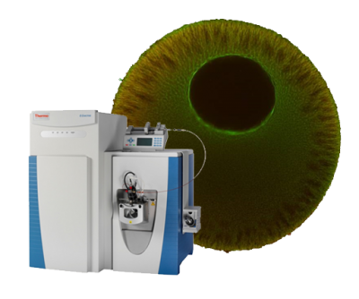
Immunofluorescence of Microtubule Assemblies in Amphibian Oocytes and Early Embryos
Publication Year
2019
Type
Journal Article
Abstract
Amphibian oocytes and embryos are classical models to study cellular and developmental processes. For these studies, it is often advantageous to visualize protein organization. However, the large size and yolk distribution make imaging of deep structures in amphibian zygotes challenging. Here we describe in detail immunofluorescence (IF) protocols for imaging microtubule assemblies in early amphibian development. We developed these protocols to elucidate how the cell division machinery adapts to drastic changes in embryonic cell sizes. We describe how to image mitotic spindles, microtubule asters, chromosomes, and nuclei in whole-mount embryos, even when they are hundreds of micrometers removed from the embryo’s surface. Though the described methods were optimized for microtubule assemblies, they have also proven useful for the visualization of other proteins.
Journal
Methods Mol Biol.
Volume
1920
Pages
17-32
Documents

